S.M. Scientific Instruments (P) LTD

3D Printing Model- Hand Blood Vessels and Nerves SMI-3DP-HVN

HomeMortuary >Virtual Dissection Table and Digital Systems >3D Printing Models >3D Printing Model- Hand Blood Vessels and Nerves SMI-3DP-HVN

3D Printing Model- Hand Blood Vessels and Nerves SMI-3DP-HVN

CLICK TO SCAN

Description

Constructing a multi-structured 3D digital anatomical model using high-precision digitized human body data. Utilizing a full-color, multi-material 3D printer and environmentally friendly resin materials to produce highly realistic 1:1 anatomical models.

Digital Human Model

The 3D-printed model of hand blood vessels and nerves is a transparent encapsulated model. The skin, superficial fascia, deep fascia, and other structures are made of transparent materials, while the bones, ligaments, blood vessels, and nerves are printed in full color. This design allows for clear observation of the internal anatomy through the transparent layers.

The internal anatomical structures are consistent with real specimens. Most of the blood vessels and nerves remain hollow, preserving their true spatial relationships and accurately representing the morphological characteristics of normal hand blood vessels and nerves. In contrast, in anatomical specimens, blood vessels and nerves often adhere to the surfaces of adjacent muscles and tendons during dissection, causing a loss of their natural anatomical positional relationships.

Data Source

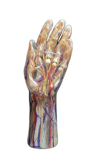
Data were selected from a high-precision digital human dataset, including raw tomographic data, refined segmentation data, and 3D geometric models of organ structures such as bones, muscles, blood vessels, nerves, ligaments, and other anatomical features. The voxel size of this dataset was 0.0384 mm × 0.0384 mm × 0.1 mm. Additionally, formalin-fixed cadaveric specimens were used as a reference for 3D printing to ensure model accuracy and facilitate comparison.

Modeling: The voxels of each anatomical structure’s surface were extracted from the original tomographic dataset to generate texture maps for the geometric model. This process ensures that the geometric model of each anatomical structure maintains the same visual perception as the real anatomical specimen.

Product Brochure

Product Brochure Link here

Featured Video